animal cell under microscope diagram
Structure of animal cell and plant cell under microscope diagrams. Light is focused on the specimen by lenses in the condensor.

Animal And Plant Cells Worksheet Inspirational 1000 Images About Plant Animal Cells On Pinterest Cells Worksheet Plant Cells Worksheet Animal Cell
Learn vocabulary terms and more with flashcards games and other study tools.

. Function cell does in the body dictate the change and adaptation done by cell. When observing onion cells there is the Cell Surface Membrane which is present in all living cells. Animal and plant cell under electron microscope.
Neuron under microscope labelled diagram. A Diagram showing the light path in a compound microscope. Most cells both animal and plant range in size between 1 and 100 micrometers and are thus visible only with the aid of a microscope.
Nuclear pore in the last lesson you. Animal Cell Diagram Under Microscope Labeled. The nucleus is covered with a rough Endoplasmic Reticulum and other organelles each designed for a specific purpose.
Throughout this article you got the different neurons labelled diagrams. Typical Animal Cell Pinocytotic vesicle Lysosome Golgi vesicles Golgi vesicles rough ER endoplasmic reticulum Smooth ER no ribosomes Cell plasma membrane Mitochondrion Golgi apparatus Nucleolus Nucleus Centrioles 2 Each composed of 9. Cell structure and organisation_notes igcsebiology dnl.
Observing a wide range of biological processes and animal cell under light microscope is easier due to advances in microscopic techniques. Under the best conditions with violet light wavelength 0. Observing a wide range of biological processes and animal cell under.
While observing with tissues or on tissue. This is due to the lack of a cell wall. The tanycytes find in the hypothalamic wall of the brains third ventricle.
Almost all animals and plants are made up of cells. Diagram 32 an animal cell. Animal cell under microscope diagram Saturday January 29 2022 Edit.
Below is the diagram of the animal cell which shows the organelles present in it. Below the basic structure is shown in the same animal cell on the left viewed with the light microscope. We all keep in mind that the human body is quite intricate and a method I.
How to see the cell nucleus under a microscope. Under the microscope animal cells appear different based on the type of the cell. Animal Cell Diagram Under Microscope Labeled.
Sunday April 18th 2021. A typical animal cell is 1020 μm in diameter which is about one-fifth the size of the smallest particle visible to the naked eye. However the internal structure and organelles are more or less similar.
We use microscope comprehensively in microbiology mineralogy cell biology biotechnology nano physics microelectronics pharmacology and forensics. Neuroglia under microscope neuron cells. The animal cell is more fluid or elastic or malleable in structure.
Unit 1 cells organ systems and ecosystems cells ppt download. A cell structure that controls which substances can enter or leave the cell. Generalized Structure of an Animal Cell Diagram You know Animal cell structure contains only 11 parts out of the 13 parts you saw in the plant cell diagram because Chloroplast and Cell Wall are available only in a plant cell.
A typical animal cell as seen in an electron microscope Medical Images For PowerPoint. Animal Cell Diagram Under Microscope. Animal cell under the microscope.
A typical animal cell is 1020 μm in diameter which is about one-fifth the size of the smallest particle visible to the naked eye. The ability to distinguish clearly the individual parts of an object under a microscope. Under the microscope animal cells appear different based on the type of the cell.
There is another modified ependymal cell tanycytes in the animals central nervous system. If you were wondering what is an animal cell below is your answer showing a picture of animal cell. One animal and one plant example given.
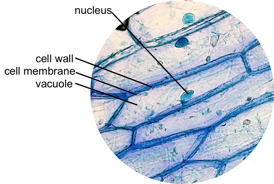
Students will observe onion cells under a microscope. When observing onion cells there is the Cell Surface Membrane which is present in all living cells. The cell is covered with cytoplasm which consists of cell organelles in it.
Plant cells have cell walls one large vacuole per cell and chloroplasts while animal cells will have a cell membrane only. This is a colored scanning electron micrograph of human red and white blood cells. Animal cells have a basic structure.
You know animal cell structure contains only 11 parts out of the 13 parts you saw in the plant cell diagram because chloroplast and cell wall are thats the major difference between plant and animal cells under microscope. The diagram below shows the general structure of an animal cell as seen under an electron microscope. A cell is the smallest functional and structural entity of life that it is easier observing animal cell under light microscope lensclutcolunch.

Year 11 Bio Key Points Cell Organelles And Their Function Cell Organelles Animal Cell Organelles

Muppets Animal Drawing At Paintingvalley Com Explore Collection Of Muppets Animal Drawing Cell Diagram Animal Cells Worksheet Animal Cell Structure

How To Draw Animal Cell Biology Drawing Animal Cell Animal Cell Drawing

Diagram Showing Anatomy Of Animal Cell Royalty Free Vector คำคมการเร ยน การศ กษา ช วว ทยาศาสตร

Animal Cell Structure And Organelles With Their Functions Animal Cell Organelles Cell Diagram

Animal Cell Organelles Cell Organelles Organelles

This Schematic Diagram Shows A Generic Animal Cell And The Organelles Including The Nucleus Endopla Human Cell Diagram Human Cell Structure Animal Cell Parts

Cells Under Electron Microscope Google Search Animal Cell Structure Animal Cells Worksheet Cell Diagram

Onion Cell Plant And Animal Cells Plant Cell Structure Plant Cell Picture

Image Result For Diagram Of Plant And Animal Cell Under Electron Microscope Celula Animal Ciencias Verdades Absolutas

Animal Cell Structure And Organelles With Their Functions Animal Cell Organelles Plant And Animal Cells

Google Image Result For Http W3 Hwdsb On Ca Hillpark Departments Science Watts Sbi3u Assigned Work Cell Struct Plant Cell Cell Diagram Animal Cells Worksheet

Animal Cell Plant Cell Diagram Cell Diagram Animal Cell

Draw It Neat How To Draw Animal Cell Animal Cell Drawing Animal Cell Cell Diagram

Epidermal Onion Cells Under A Microscope Plant Cells Appear Polygonal From The Cell Diagram Plant Cell Diagram Plant Cell

Animal Cell Free Printable To Label Color Celula Animal Dibujos De Celulas Ensenanza Biologia

Plant Cell Diagram Animal Cell Diagram Plant And Animal Cells Science Cells Animal Cell

Learn About The Plant Cell Science For Kids And Science Activities And Projects For Kids Plant Cell Cell Diagram Animal Cell Structure
